


A newly developed imaging technique has led to a surprising discovery in the world of microbiology. A research team led by Ralph Youngmann at the Max Planck Institute (MPI) for Biochemistry and at the Ludwig Maximilian University (LMU) participated, along with Eugenio F. Fornasero and Felipe Opazo, who work as working group leaders at the University Medical Center. Göttingen (UMG), and the Helmholtz Center in Munich. A high-throughput spatial imaging method called SUM-PAINT enabled them to develop a neuronal atlas with single-molecule resolution. They have discovered a previously unknown type of synapse. The researchers presented their findings in a study conducted by Edward Untrauer in Youngman's laboratory. It appeared in issue No. 187/7 of the specialized magazine “Cell” (open access articlePDF available). The SUM-PAINT process described here includes a data generation and analysis workflow that can be used by researchers around the world. As the study notes, it can be used relatively easily with standard microscopes and does not require any special equipment.
advertisement
Jungmann is convinced that SUM-PAINT is not just a milestone on the way to unraveling the complexity of cell biology at the molecular level. The method also has what it takes to enable a breakthrough in the discovery of new therapeutic approaches for neurodegenerative diseases. It provides the opportunity to investigate previously hidden details of neurological disorders and can thus contribute to a deeper understanding of the underlying mechanisms of diseases such as Parkinson's disease or Alzheimer's dementia.
Synapses as complex protein structures
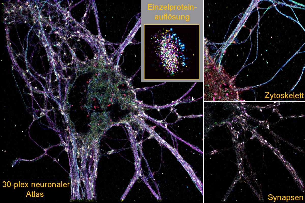
For the first time, SUM-PAINT makes it possible to display and map a large number of proteins simultaneously with molecular resolution and at lightning speed. As Untrauer explains, one must simultaneously examine the location, identity, and interaction of individual biomolecules in order to understand the complexity of living systems down to the smallest level. There are four critical challenges to overcome: In addition to the ability to combine multiple signals, this concerns the criteria of sensitivity, throughput and spatial resolution. The team looked at the complex environment of neurons in the human brain and, for the first time, developed a neural atlas with single-molecule resolution of 30 different types of proteins. He was able to decipher the complexity of the synaptic protein composition of nearly 900 individual synapses. This resulted in large amounts of data – which in turn could only be examined in detail with the help of a specially developed analysis pipeline powered by machine learning. The analysis captured 1,600 features from the imaging datasets. Characteristics included protein content, distribution, and shape. The researchers discovered a previously unknown type of synapse during this analysis. It makes up only about one percent of all synapses in the human brain. It was not detectable using other imaging techniques.
This study, published on an open access basis, allows interested parties worldwide to implement highly detailed imaging for their own purposes based on the described method.
(PSZ)

“Prone to fits of apathy. Zombie ninja. Entrepreneur. Organizer. Evil travel aficionado. Coffee practitioner. Beer lover.”
More Stories
AOC Graphic Pro U3: A new color-accurate monitor series for creative people
Do you already know Ruona? -Dukchik
The “Dragon” must move to the International Space Station